Near infrared light is light with a wavelength of 800 nm to 2500 nm, and
absorption and scattering of light in the living body are smaller than visible light, and it is possible
to observe deep parts of the living body that can not be observed with the naked eye . Therefore, near infrared light is
actively used in the medical field. Among medical equipment using near-infrared light, in this research,
we focused on developing an endoscope using near-infrared light. Endoscopes are non-invasive and do not need
to be hurt by a scalpel and can be observed in real time.
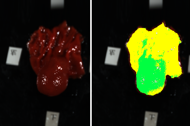
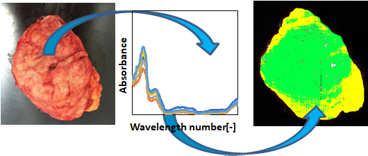
In this study, it is difficult to detect visually and is difficult to detect early in the muscular layer
in the submucosa of digestive organs such as the stomach and small intestine, and it is difficult to detect early and
to the digestive tract called GIST (interstitial stromal tumor). The research target is a tumor that can be
When making a diagnosis, use machine learning to determine whether each cell is a tumor cell or a normal cell, and
the tumor cells are drawn in green and normal cells are drawn in yellow. When creating learning data used in machine learning,
marking by a doctor is used.
Close
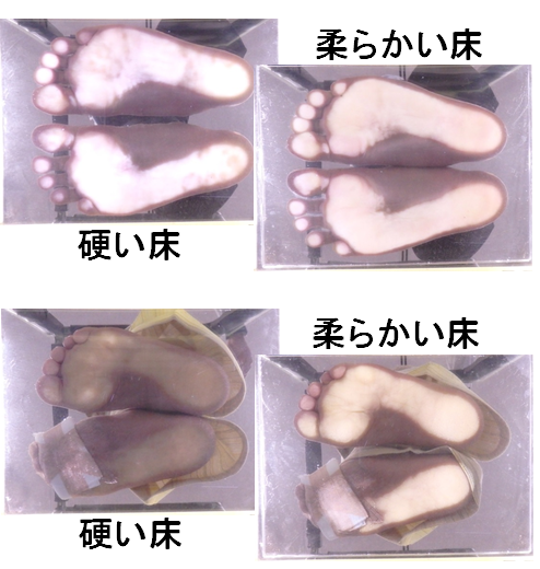
In present-day Japan, there are more than one in 10 patients suspected of having diabetes.
Furthermore, 1 in 3 people suffer from a disease called diabetic neuropathy. If the disease gets worse,
it will lead to necrosis and amputation of the foot, treatment is extremely difficult, but if the symptoms are light,
treatment with medical treatment etc. is possible.
However, current inspection methods require a great deal of work. Therefore, it is expected to establish a method that
enables early detection of diabetic neuropathy by a different examination method from the past. Previous studies
have shown that diabetic neuropathy patients have poor blood flow in the foot and that the amount of sweating reduces
the skin on the sole of the foot.
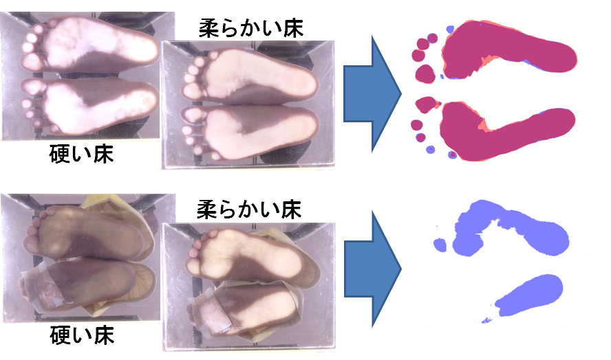
In this research, we think that differences occur in the contact area of the sole and floor on soft and hard floors, and
propose a method to determine the severity by comparing the magnitude of the difference from image processing did.
Currently we are conducting experiments to verify the usefulness of the proposed method.
Close
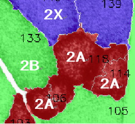
Skeletal muscle weakness is an important factor in many different chronic diseases and aging,
so it is an important task to elucidate the skeletal muscle weakness mechanism nowadays
when the improvement of healthy life span is required. Important morphological features of muscle fibers,
such as the cross-sectional area, shape, type, number and location of muscle fibers, are one of the important factors
that determine muscle health and function. Histological analysis of skeletal muscle in consideration of these morphological
features is required to elucidate the relationship between chronic disease and aging and skeletal muscle weakness.
However, at present, quantification of the morphological features of these muscles is still a manual or
semi-automatic method based on muscle measurements and requires extensive human intervention. It is required
to analyze skeletal muscle to reduce the time and effort required to quantify the morphological characteristics of muscle.
Therefore, we developed software that automates the task of image analysis of skeletal muscle
by utilizing image processing.
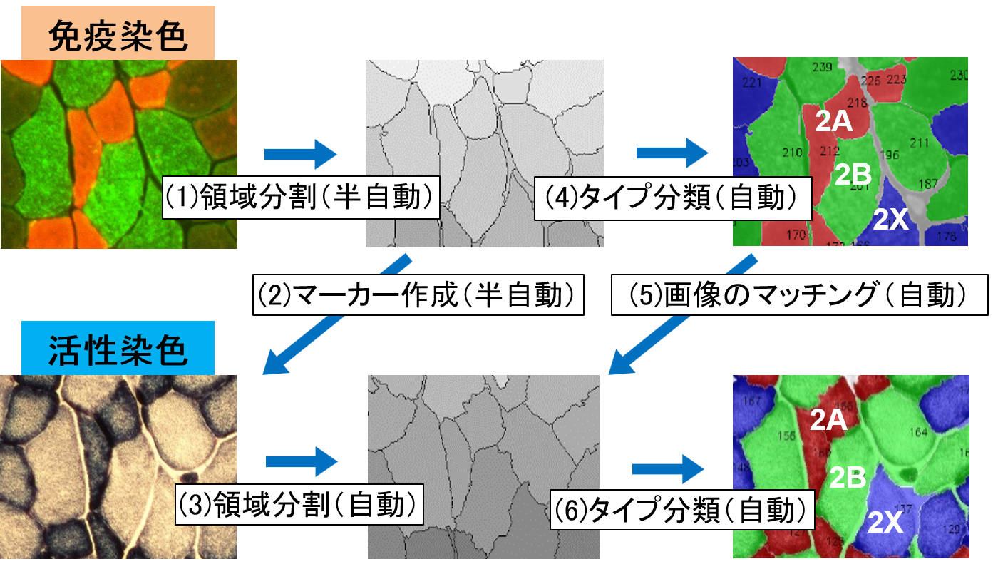
Apply γ correction as pre-processing to the Watershed method so that the cell division in the image
can be divided into regions simply by one-touch with the mouse, and to classify the cells by type,
the luminance value for each cell or Automatic classification was made possible from the values of color and
cross-sectional area independently of the human eye. As a result,
it became possible to reduce the analysis time to 1/5.
Close